Tarih: Issue 65 - February 2016
Introduction
Unfortunately, we currently come across more gunshot wounds (GSW) due to military conflict, increasing range of armed attacks, suicide cases, terrorist attacks towards civilians and troops. In certain societies, the number of GSW cases exceed the injuries related with motor vehicle accidents. In the United States of America, where usage of weapons is extensive, the annual number of nonfatal GSW cases is around 100,000. (1)
Ballistic Impact
The rate of destruction varies due to the mass, caliber, speed, groove of the bullet/particle. The firing distance is another factor affecting the impact of the bullet. The penetration power of the bullet/particle that enters the body is closely related with the amount of its kinetic energy. The penetration capacity and lethalness of the bullet/particle increase in parallel with its speed. (2,3,4). The destruction capacity of the bullet also changes when the bullet/particle changes its direction as it hits the bone in the trajectory and when it loops. The particle effect of the broken bone alters the ballistic impact of the bullet as well.
Introduction to Medical Technology
Following the first response to the injured patient as soon as they enter the hospital and after they overcome the trauma, in order to let themreturn to daily life and to feel better psychologically, it is of vital importance that the injured body part is repaired back to its former functional and cosmetic structure. At this stage, during the treatment of the injured bone structure, in addition to the patient’s own bone tissue, implants designed especially for that patient are utilized. The period during which the patient is anesthetized is decreased when the operation simulation is conducted before narcosis and meanwhile the locations of the bone segments are identified, screws are selected and positioned beforehand. This decreases the duration of the operation as well as the rate of failure, thus increases the success of the operation. According to research, every hour under the influence of narcosis delays the patients’ adaptation to previous daily life for one additional day.
3D Imaging of Patients
Through the developed and highly sensitive 3 Dimensional Computer Tomography (CT) devices, the internal organs of the patients can be imaged accurately (DICOM, TIFF, Interfile, GIF, JPEG, PNG, BMP, PGM, MRC, RAW). The special software used in this area segment the tomographic scans and converts them into the format utilized by the operation simulation, 3D medical model devices and design software (.stl). Via this software, the 3D images of even the stones and tumors emerging in the bones, veins and muscular tissues can be identified with their relevant positions depending on their level of density.
3D Medical Models
The 3D models of the positions of the bones and tissues relation to each other are used by the surgeon,for patient communication and to predeterminefaultles implant lines. Building a clear communication with the patient, informing the patient, making precise decisions for the treatment of the patient and perfect compatibility of the implant with the patient result in the rapid preparation of the patient for the surgery, quicker recovery and accelerates the adaptation of the patient to future life. Three-dimensional physical models are quite valuable assets, both for surgeons and surgical patients nowadays. Identification of the screw orbits, screw selection, selection of the surgical devices and technical operations can be conducted through these models.
Planning Surgeries on Models
Dislocated bones, due to trauma, are brought to the position where they should be through the related software interfaces as a result of the studies conducted jointly by the relevant doctors and engineers. Meanwhile the softer elements such as the veins, muscles and connective tissues are imaged and studied. Since an implant applying pressure to the vein, a screw coinciding with a nerve or an implant that cannot be located to its place due to the connective tissue should be avoided, these points must be taken into consideration. Moreover, through this software; distance, angles, volume and density can be measured.
On Bone Structure
The bone is divided into two parts on a macro level; the cortical (or compact) and cancellous (or trabecular) bone. These two structures are different from each other in their density and porosity. The micro variations in density cause significant changes in the endurance and elasticity module values. The cancellous (trabecular) bone is composed of bone material in the form of a short stick known as the trabecula and has a spongy look. Cancellous bone is mostly situated at the inner surface of the cortical bone and at the ends of the long bones. Both bones are basically composed of the same material. The irregular yet optimized arrangement and orientation of the components results in the heterogeneous and anisotropic bone material. The skeleton system is a structure protecting the visceral organs while creating a durable kinematic link, providing the muscles a point for adherence and eventually supporting the body movements. The biomechanics of the bone is specialized in order to conduct such complex tasks. The bone is a tissue that is capable of auto-repairing and adapting its internal structure and configuration in accordance with the varying mechanic requirements. Long-standing overload or underload definitely alters the bone density. Compared with iron, bone is three times lighter and ten times more flexible. The collagen content is in charge of resisting against contortion, while mineral content applies resistance against the pressure. The significant terms herein are endurance and rigidity. (5)
Using Patient’s Own Bone Tissue
During the restructuring of the bone, treatments through using the patient’s own bones (flaps) extracted from other parts of the body can be executed. The flap is removed from the places of the tissue particle fed from the named or anonymous blood veins in the body, and used for providing tissue that is missing in a remote or close part of the body. The flap tissue is transported to another part,either without pausing blood circulation or repaired in the place where it is removed, and fed through the veins at that location.
As they are thicker, bigger and since numerous tissues could be simultaneously used for transportation, the flaps are applied for the treatment of wide, deeper and more complex injuries. The skin, muscle, bone and fascia tissues can be used individually or collectively. The most important difference of the flaps from the grafts is the existence of the aorta and/or nerves in the flaps. Tissues such as the dermis, subcutaneous tissue, muscle, bone, cartilage, nerve, vein, tendon, and fascia can be located either individually or collectively (6).
Within this scope, during the full placement, osteotomy, puncturing and referencing of the flap removed from the patient, surgical guides are used. Surgical guides are guiding parts that are designed in order to increase the surgical success and for shortening duration of the surgery, for abolishing the risks that may occur during the surgery and that allow the exact application of the 3D and computer aided surgical planning, usually manufactured with polymer or sometimes from metal. The production of the guides is accomplished with maximum 0.1 precision rate through developed 3D printers. They allow sterilization with ethylene oxide or autoclave. They do not react with the body in any way, they are designed for single use and they could not be left in the body (7).
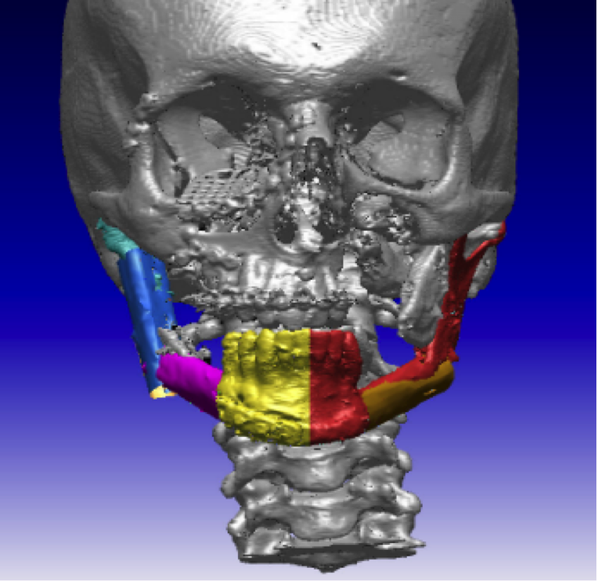
Example of the Surgical Guide Designed for the Application of the Fibula Bone in Reconstruction of Mandibula.
Guides are designed with anatomical compatibility; they are perfectly located on the bone and fixed from their relevant parts to the given reference points so that they do not move during the osteotomy or puncturing. They are designed with the joint efforts of the doctors and engineers in a computer environment for the surgery simulation results, executing accurate, faultless incisions and punctures.
The principal advantages of this method; as fewer incisions are conducted less bone tissue is lost. As a result of the planned incision sections, full contact is accomplished between the bone segments and the healing – ossification and stability would be improved. Since the surgery planning is conducted beforehand, the surgery and as the issues that may arise can be foreseen, the duration of the surgeries are shortened, the fault rate is decreased and the success rate increases (8).
Patient Specific Implant Design
The detailed imaging of the visceral organ via the high-technology imaging devices (CT, MR), converting this image to operable details via computer aided designs, taking the biocompatible material’s characteristics in addition to the anatomic structure and bone features into consideration, and thus solving the existing problem of the implant, designing and developing the solution without harming the tissues, are required.
The methodology is generally as follows;
Before the design, the detailed tomography of the diagnosed patient who lost a bone limb is taken.
The captured section images are united through a computer-aided study, and the existing bone loss, the losses endured until the surgery if the loss is caused by cancer disease, the part to be removed during the surgery are determined and plans as well as related information are identified by the relevant doctors and engineers.
Bone density measures are executed for all the bones that the implant will relate with.
The weight, age and biomechanical movements of the patients are important points to be taken into consideration.
Length, angle and area measurements are conducted over the image.
The designs compatible with the bone structure and convenient for the locations where bone loss occurs are executed based on the acquired 3D data. These designs obtained through a point cloud could be accomplished by getting surfaces and installing reference planes and curves from available locations as they could also be realized with systems allowing flexibility. In addition to biologic compatibility, the endurance of the implant is the most essential requirement. For instance, in the fractured spots where the soft tissue thickness is less, a bone implant’s distracting the tissues from the bone can damage these tissues and cause complications. If the implant is designed for high mechanic endurance and fatigue resistance, it can be too rigid and that will result in the deficiency of the bone in enduring the physiological loads and in poor reshaping of the bone. Considering the implantation and the selection of the implant material, the mechanic and physical features of the bone must be perceived well. Between the implant material and the surrounding tissue, there are always mechanic and biological interactions. The resistance between the implant and the bone depends on this interaction. The rigidity of the screw and the implant utilized during the fixation must be proportioned to each other and to the bone, if not the screw’s tooth may be scraped from the bone or the screw may be broken.
Analysis
Throughout their utilization period, the implants and prosthetics designed in alignment with the human anatomy, must resist the loads. The interaction with the bone and the selected screws should be foreseen, the impact of fatigue and corrosion on the lifetime of the implant and prosthetic must be well calculated. The implant and prosthetics inside the body should be used in environments with hyper-variable conditions in the body. Average load on a single hip joint may increase up to three times of the body weight. During activities such as jumping, this value may be tenfold of the body weight. This stretching of our body constantly repeats itself during activities such as standing, chewing, sitting and running.
Manufacturing the Implant Through 3D Technology
At this stage of the material and manufacturing technology development, titanium (Ti) alloy powders are combined in layers via high technology and form complex geometries in pored and dense structures (sintering in gas environment between Ti6Al4V 1100 ºC-1350 ºC temperatures). With sintering, as a result of growing particle contact points the specific surface area shrinks, decrease in bore density or its globalization is enabled, moreover the atom spaces that may occur in the structure or crystal faults such as dislocation are decreased.


.jpg)